
Item no.: P2538000
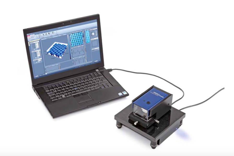
Principle
Approaching a sharp silicon tip mounted on a cantilever to a sample surface leads to an atomic scale interaction. The result is a bend of the cantilever which is detected by a laser. In static mode the resulting deflection is used to investigate the topography of the sample surface line-by-line using a feedback loop. In dynamic mode the cantilever is oscillated at fixed frequency resulting in a damped amplitude near the surface. The measurement parameters (setpoint, feedback gain,…) play a crucial role for image quality. The dependence on the imaging quality is investigated for different nano structured samples.
Tasks
- Set-up the microscope and start up the software. Mount a cantilever (with tip) and approach the tip towards a sample.
- Investigate the influence of the scanning parameters on the imaging quality and performance, e.g. PID gain, setpoint (force), vibrational amplitude, and scanning speed. Use both static and dynamic force mode.
- Image 7 different samples (microstructures, carbon nano tubes, skin cross-section, bacteria, CD stamper, chip structure, glass beads) by optimizing the parameters respectively.
What you can learn about
- Atomic Force Microscopy (AFM)
- Lennard-Jones potential
- Imaging of nano structures
- Static Force Mode
- Dynamic Force Mode
- Feedback loop
- Force
- Vibrational amplitude
Software included. Computer not provided.