
Item no.: R02/1 [1013869]
- Weight: 1.7 kg
- Dimensions: 60 x 40 x 6 cm
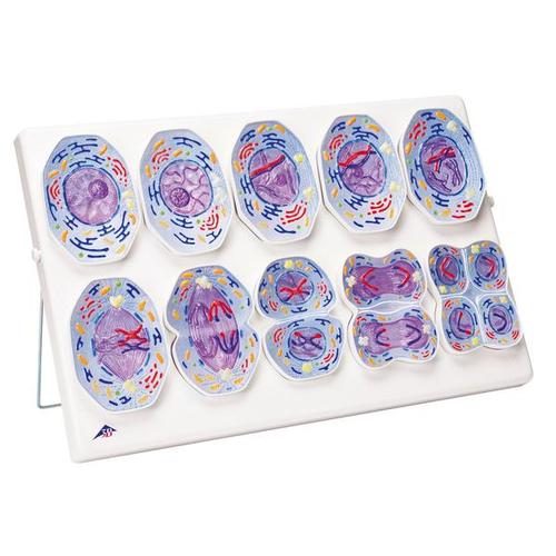
This newly developed 3B Scientific® meiosis model series shows the 10 stages of meiosis on the basis of a typical mammal cell at an enlargement of approximately 10,000 times. The detailed stages of meiosis:
1. Interphase (stage of G1-phase)
2. Prophase I (leptotene)
3. Prophase I (zygotene and pachytene)
4. Prophase I (diplotene)
5. Prophase I (diakinesis)
6. Metaphase I
7. Anaphase I
8. Telophase I, cytokinesis I, interkinesis, prophase II and metaphase II
9. Anaphase II
10. Telophase II and cytokinesis II
The three-dimensional relief models are painted according to the usual coloring methods of microscopy, making the process of meiotic cell division easy to understand. The cell organelles are portrayed open in the lower part of the models.
The meiosis models are equipped with magnets at the rear so that they can be easily arranged on a magnetic board in the classroom. The model series is supplied in a storage system (40 x 60 cm) that can be hung on a wall. The meiosis model comes complete with a detailed description and handouts to copy for your lessons.